Renovascular Conditions India offers information on Renovascular Conditions in India, Renovascular Conditions cost India, Renovascular Conditions hospital in India, Delhi, Mumbai, Chennai, Hyderabad & Bangalore, Renovascular Conditions Surgeon in India
What are renovascular conditions?
 Renovascular conditions affect the blood vessels of your kidneys, called the renal arteries and veins. When the blood flow is normal through your kidneys, your kidneys rid your body of wastes. The kidneys filter these wastes into your urine, which collects in your bladder, and from there the wastes exit your body when you urinate. Your kidneys also help control your blood pressure by sensing the blood pressure and secreting a hormone, called renin, into your bloodstream. The amount of renin secreted by your kidneys can help regulate your blood pressure if it is too high or too low. When your kidney blood vessels narrow or have a clot, your kidney is less able to do its work. Your physician may diagnose you with renal artery stenosis or renal vein thrombosis.
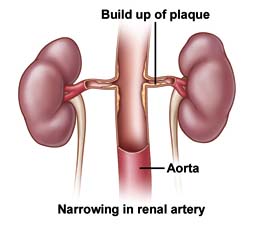
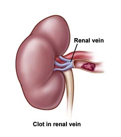
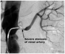
Renal artery stenosis is the narrowing of kidney arteries. This condition may cause high blood pressure and may eventually lead to kidney failure. Renal vein thrombosis means that you have a blood clot blocking a vein in your kidney. Blood clots in renal veins are uncommon and rarely affect the kidney, but they can sometimes travel to and lodge in arteries supplying your lungs, causing a dangerous condition called a pulmonary embolism.
 What are the symptoms?
You may not notice any symptoms. Renovascular conditions develop slowly and worsen over time. If you have high blood pressure, the first sign that you may have renal artery stenosis is that your high blood pressure may become worse or the medications that you take to control your high blood pressure may not be as effective. Other signs of renal artery stenosis are a whooshing sound in your abdomen that your physician hears through a stethoscope, decreased kidney function, congestive heart failure or, eventually, a small shrunken kidney.
When renal vein thrombosis occurs, a clot in your vein may break free or block the flow in a healthy blood vessel. If this happens, symptoms may include :
- Pain in the sides of your abdomen, legs, or thighs
- Blood in your urine
- Protein in your urine
- An enlarged kidney that your physician can feel
- Fever, nausea, or vomiting
- High blood pressure
- Sudden, severe swelling in your leg
- Difficulty breathing.
 What causes renovascular conditions?
Hardening of the arteries causes renal artery stenosis. Your arteries are normally smooth and unobstructed on the inside but, as you age, a sticky substance called plaque can build up in the walls of your arteries. Cholesterol, calcium, and fibrous tissue make up this plaque. As more plaque builds up, your arteries can narrow and stiffen. This is the process of atherosclerosis, or hardening of the arteries. Eventually, enough plaque may build up to interfere with blood flow in your renal arteries.
Smoking, obesity, advanced age, high cholesterol, diabetes, and a family history of cardiovascular disease are factors that may increase your chances for developing atherosclerosis.
Nephrotic syndrome is the most common cause of a clot in the renal vein (renal vein thrombosis). Nephrotic syndrome is a condition in which large amounts of a protein called albumin leak into your urine. Other causes of renal vein thrombosis include injury to the vein, infection, or a tumor.
What tests will I need?
Your physician will recommend the following tests to help determine if you have renal artery stenosis :
- Ultrasound, which allows your physician to see your blood vessels and organs using high-frequency sound waves. Using ultrasound, your physician can locate clots or narrow areas inside your arteries and also determine the size of your kidney
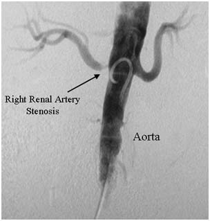
- Angiography, which is more invasive, can locate a narrowing or blockage, can measure blood flow, and can be used to sample blood for testing. In this test, your physician injects a contrast dye into your vessels through a small catheter and then takes x-rays. The structure inside of your vessels appears on the x-ray because x-rays cannot pass through the dye. This test finds the location and pattern of blockages in your kidney blood vessels. The dye itself is later excreted through your kidneys and it sometimes can affect their function
- Spiral computed tomography (CT) scan, which creates detailed three-dimensional images from x-rays of slices of your body. This study also sometimes uses contrast dye
- Magnetic resonance angiography (MRA), which uses magnetic fields and radio waves to produce three-dimensional images of your arteries. This study also sometimes uses contrast dye
- Radionuclide scanning, which uses a radioactive substance and a special camera to analyze the blood flow through your kidney.
If your physician suspects you may have renal vein thrombosis, he or she may perform a Doppler ultrasound. Doppler ultrasound uses short bursts of high-frequency sound waves to create real-time, moving pictures of blood flowing through your vessels. If the Doppler ultrasound reveals a possible clot, your physician may use CT scans, MRA, or venacavography to further locate it. Venacavography creates pictures of your main abdominal veins. It is a form of angiography during which your physician will inject a contrast dye to view the blood flow through your veins on an x-ray monitor.
How are renovascular conditions treated?
Medication
If your physician diagnoses renal artery stenosis, he or she may prescribe blood pressure medications. Some medications may include:
- Diuretics
- Beta-blockers
- Angiotensin-converting enzyme (ACE) inhibitors
- Calcium channel blockers.
Thrombolysis
If you experience sudden blockage in your renal artery, your physician may recommend a procedure called thrombolysis. In thrombolysis, a vascular physician injects a clot-dissolving medication directly to a clot through a long, thin tube called a catheter. This procedure, when needed, is often done at the time of angiography.
If your physician diagnoses renal vein thrombosis, he or she may give you anticoagulants. These medications decrease your blood's ability to clot. In critical cases of renal vein thrombosis, your physician may perform thrombolysis.
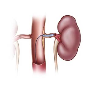
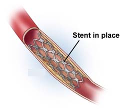
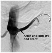
Angioplasty and stenting
If your renal artery is partially or completely blocked, your physician may recommend a procedure called angioplasty and stenting. To perform this procedure, your physician inserts a catheter through a small puncture site, or sometimes a small incision, and guides it through your blood vessels to your renal artery. The catheter carries a tiny balloon that inflates and deflates, flattening the plaque against the walls of your artery. Next, your physician may insert a tiny metal-mesh tube called a stent in the artery to hold it open. This procedure, when needed, is often performed at the time of angiography.
Related Pages >>
 Stent Placement Surgery
Surgery India Renovascular Conditions, Cost Renovascular Conditions, Radiological Diagnosis And Treatment, Renovascular, India Kidney, India Artery Stenosis, India Vein Thrombosis, India Smoking, India Angiography, India CT Scan, India MRA, India Medication, India Thrombolysis, India Angioplasty And Stenting, India Endarterectomy, India Bypass Surgery, India Renovascular Conditions, India Cost Renovascular Conditions Treatment, India Carotid Renovascular Conditions Treatment, India High Blood Pressure, India Pulmonary Embolism, India Blood Clot, India Hardening Of The Arteries, India Atherosclerosis, India Nephrotic Syndrome, India High Cholesterol, India Low Cost Renovascular Conditions Treatment Delhi, India Renovascular Conditions Treatment Hospital Delhi, India Renovascular Conditions Treatment Mumbai Hospital, India Cost Renovascular Conditions Treatment Mumbai